
Floaters—those tiny specks, strings, or cobweb-like shapes that drift across your field of vision—are more common than many realize. Most people experience them at some point, especially as they age. While often harmless, floaters can sometimes signal serious eye conditions. Understanding what causes them, recognizing when they might indicate a problem, and knowing when to seek professional evaluation are essential steps in preserving long-term eye health.
What Are Floaters and How Do They Form?
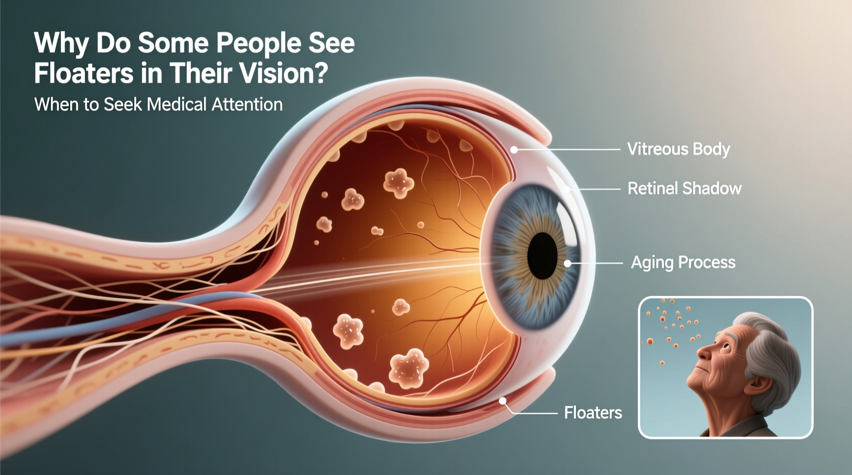
Floaters are small deposits or clumps of collagen within the vitreous humor—the clear, gel-like substance that fills the inside of the eye. As light passes through the eye, these particles cast shadows on the retina, which appear as floating spots or strands in your vision.
The vitreous is 99% water and 1% collagen fibers arranged in a lattice structure. With age, this gel begins to liquefy and shrink—a process called vitreous syneresis. As it pulls away from the retina, microscopic fibers can clump together, forming what we perceive as floaters.
This natural aging process typically begins in adulthood, often between ages 50 and 70, but can occur earlier in nearsighted individuals or those who’ve had eye trauma or surgery.
Tip: Floaters are usually more visible against bright, uniform backgrounds like white walls or clear skies.Common Causes of Eye Floaters
While aging is the most frequent cause, several other factors contribute to the development of floaters:
- Aging-related vitreous changes: The most common reason, especially after age 50.
- Nearsightedness (myopia): People with high myopia have longer eyeballs, increasing stress on the vitreous and accelerating its degeneration.
- Posterior vitreous detachment (PVD): A condition where the vitreous separates from the retina. It affects over 75% of people over 65 and often causes a sudden increase in floaters.
- Eye inflammation (uveitis): Inflammation inside the eye can release cellular debris into the vitreous.
- Eye injury or trauma: Blunt force or surgical procedures may disrupt the vitreous structure.
- Diabetic retinopathy: Abnormal blood vessel growth and leakage can introduce particles into the vitreous.
- Retinal tears or detachments: More serious conditions that may present with new floaters alongside flashes of light.
“Floaters are usually benign, but a sudden shower of new ones should never be ignored. They can be the first sign of a retinal tear.” — Dr. Lena Torres, Retina Specialist, Massachusetts Eye Institute
When Floaters Signal a Medical Emergency
Most floaters are not dangerous and tend to become less noticeable over time as the brain adapts. However, certain symptoms require immediate ophthalmologic evaluation.
A sudden appearance of dozens of new floaters, particularly if accompanied by flashing lights or a curtain-like shadow over part of your vision, could indicate a retinal tear or detachment—a sight-threatening emergency.
The retina is the light-sensitive layer at the back of the eye. If it detaches from its underlying tissue, permanent vision loss can occur within hours without treatment. Early detection dramatically improves outcomes.
Symptoms That Require Immediate Evaluation
| Symptom | Possible Cause | Action Required |
|---|---|---|
| Sudden onset of many new floaters | Posterior vitreous detachment, retinal tear | See an eye doctor within 24–48 hours |
| Flashes of light (photopsia) | Vitreous tugging on retina | Urgent evaluation needed |
| Curtain or shadow in peripheral vision | Retinal detachment | Emergency care—go to an ophthalmologist or ER |
| Blurred or distorted central vision | Macular involvement or swelling | Prompt assessment required |
| Pain with visual changes | Inflammation or infection | Seek care immediately |
Real-Life Scenario: When Ignoring Floaters Had Consequences
Mark, a 58-year-old graphic designer, began noticing a cluster of new floaters in his right eye one Monday morning. He dismissed them, assuming they were due to fatigue. Over the next two days, he experienced flickering lights in his peripheral vision and a growing dark area on the left side of his visual field. By Wednesday, his vision was significantly impaired.
He visited an ophthalmologist, who diagnosed a large retinal detachment. Surgery was performed urgently, but because treatment was delayed, Mark lost some peripheral vision permanently. His doctor emphasized that if he had come in within 24 hours of symptom onset, the outcome might have been far better.
This case illustrates how quickly a seemingly minor visual disturbance can escalate into irreversible damage. Awareness and timely action are critical.
How Eye Doctors Diagnose the Cause of Floaters
When you report persistent or new floaters, an eye care professional will conduct a comprehensive dilated eye exam. This involves using special drops to widen the pupils, allowing a clear view of the vitreous and retina.
During the exam, the doctor uses an ophthalmoscope or slit lamp to inspect for:
- Signs of posterior vitreous detachment
- Retinal tears or holes
- Areas of thinning or detachment
- Bleeding or inflammatory cells in the vitreous
In some cases, optical coherence tomography (OCT) or ultrasound imaging may be used to obtain detailed cross-sectional views of the retina, especially if the view is obstructed by cataracts or blood.
Step-by-Step: What to Expect During a Floater Evaluation
- Medical history review: Discuss symptoms, onset, duration, and any risk factors like diabetes or prior eye injuries.
- Visual acuity test: Measure clarity of vision with an eye chart.
- Pupil dilation: Administer drops to enlarge the pupils (takes 15–30 minutes).
- Dilated fundus examination: Use specialized lenses to examine the retina and vitreous.
- Peripheral retinal screening: Look for tears, holes, or signs of detachment.
- Imaging (if needed): OCT or ultrasound to assess structural integrity.
- Diagnosis and plan: Determine whether observation, laser treatment, or surgery is necessary.
Treatment Options for Problematic Floaters
Most floaters don’t require treatment and fade into the background over months or years. However, when floaters severely impair vision or quality of life, interventions may be considered.
1. Observation and Adaptation
The majority of patients adapt neurologically to floaters. The brain learns to filter them out during normal visual processing, making them less intrusive over time.
2. Laser Vitreolysis
In select cases, an ophthalmologist may use a YAG laser to break up large, bothersome floaters. This outpatient procedure targets specific opacities in the vitreous, vaporizing them into smaller, less noticeable fragments.
Success varies, and risks include retinal damage or increased intraocular pressure. It’s not widely recommended as a first-line treatment.
3. Vitrectomy Surgery
For severe cases, a pars plana vitrectomy removes the vitreous gel entirely and replaces it with a saline solution. This eliminates floaters but carries significant risks, including cataract formation, retinal detachment, and infection.
Vitrectomy is typically reserved for patients with debilitating symptoms and no improvement over time.
“We only consider vitrectomy when floaters are truly disabling and all other options have failed. The risks must be carefully weighed.” — Dr. Alan Zhou, Vitreoretinal Surgeon, Johns Hopkins Medicine
Frequently Asked Questions About Floaters
Can floaters go away on their own?
Yes, many floaters become less noticeable over time. They may settle below the line of sight or be absorbed by the eye. The brain also adapts by suppressing awareness of persistent visual noise. However, they rarely disappear completely.
Are floaters a sign of blindness?
Not directly. Floaters themselves do not cause blindness. But if they are associated with retinal tears or detachment, untreated complications can lead to permanent vision loss. Prompt diagnosis is key to preventing such outcomes.
Should I worry about floaters if I’m under 40?
Floaters can occur at any age, especially in people with high myopia, diabetes, or a history of eye injury. While less common in younger adults, new floaters still warrant evaluation to rule out underlying issues like inflammation or early PVD.
Preventive Care and Eye Health Checklist
While you can’t prevent all floaters, especially those tied to aging, proactive eye care reduces the risk of complications. Use this checklist to stay ahead:
- ✅ Schedule regular eye exams—at least every two years, or annually if over 60 or at higher risk.
- ✅ Report any sudden change in vision, including new floaters or flashes, immediately.
- ✅ Manage systemic conditions like diabetes and hypertension that affect eye health.
- ✅ Wear protective eyewear during sports or DIY projects to prevent trauma.
- ✅ Avoid smoking, which increases oxidative stress and accelerates eye aging.
- ✅ Maintain a diet rich in antioxidants—leafy greens, fish, nuts, and colorful vegetables support retinal function.
Conclusion: Don’t Ignore Changes in Your Vision
Floaters are a common part of the eye’s aging process and usually pose no threat. But when they appear suddenly, multiply rapidly, or accompany flashes and vision loss, they demand urgent attention. The difference between preserving full vision and suffering permanent damage can hinge on just a few hours.
Your eyes provide a window into both your ocular and overall health. Conditions like diabetes, hypertension, and autoimmune diseases often manifest first through changes in the eye. Regular monitoring isn’t just about clarity—it’s about catching problems before they escalate.
🚀 If you’ve noticed new or worsening floaters, don’t wait. Schedule a comprehensive eye exam today. Your vision is worth protecting—one floater at a time.