
The human eye is a marvel of biological engineering, and one of its most noticeable features is the stark contrast between the colored iris and the surrounding white surface. This white portion—commonly referred to as the \"white of the eye\"—plays a crucial role in both structure and function. But why exactly are eyeballs white? What purpose does this white layer serve? And what does it mean when that whiteness changes? Understanding the anatomy, physiology, and potential issues related to the sclera provides valuable insight into ocular health and overall well-being.
Anatomy of the Sclera: The Eye’s Protective Shell
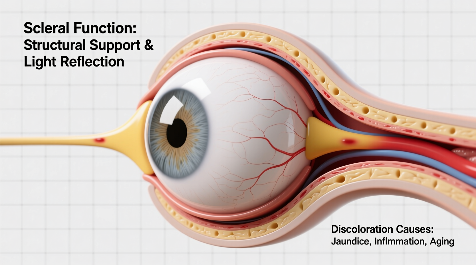
The white part of the eye is called the sclera. It makes up about 80% of the outer coat of the eyeball and extends from the cornea at the front to the optic nerve at the back. Composed primarily of dense connective tissue rich in collagen and elastic fibers, the sclera is tough, fibrous, and opaque—qualities essential for protecting the delicate internal structures of the eye.
Beneath the conjunctiva—a thin, transparent membrane that covers the sclera—the surface appears smooth and white under normal conditions. This appearance is due to the way light scatters through the densely packed collagen fibers, which lack pigmentation. Unlike the skin or other tissues, the sclera does not contain melanocytes (pigment-producing cells), contributing to its pale color.
“Structurally, the sclera acts like a protective helmet for the eye, maintaining shape and shielding against trauma.” — Dr. Lena Patel, Ophthalmologist and Clinical Researcher
Functions of the Sclera Beyond Whiteness
The sclera's whiteness is more than just cosmetic—it reflects its functional design. Key roles include:
- Mechanical Protection: Shields the retina, lens, and vitreous humor from physical injury.
- Structural Support: Maintains the spherical shape of the eyeball, critical for proper focusing of light.
- Attachment Site for Extraocular Muscles: Six muscles responsible for eye movement anchor directly into the sclera, enabling precise control over gaze direction.
- Barrier Against Pathogens: Its thickness and avascular nature (low blood supply) reduce vulnerability to infection.
In addition, the sclera helps regulate intraocular pressure by resisting deformation. Any weakening or thinning can lead to serious conditions such as ectasia or staphyloma, where the eye bulges abnormally.
Tip: Protect your sclera—and overall eye health—by wearing UV-blocking sunglasses outdoors. Prolonged sun exposure can cause degenerative changes in the scleral tissue.Common Causes of Scleral Discoloration
While a bright white sclera is typically a sign of good health, discoloration can signal underlying issues. Yellowing, redness, blueness, or patchy pigmentation should not be ignored. Below are common causes categorized by color change:
| Discoloration | Possible Causes | Associated Symptoms |
|---|---|---|
| Yellow | Hepatitis, cirrhosis, jaundice (elevated bilirubin) | Fatigue, dark urine, abdominal pain |
| Red/Pink | Conjunctivitis, subconjunctival hemorrhage, uveitis | Irritation, discharge, light sensitivity |
| Blue/Grey | Thin sclera (genetic or age-related), osteogenesis imperfecta | Translucent appearance, fragile bones (in syndromic cases) |
| Dark Spots/Brown Patches | Primary acquired melanosis, nevi, racial melanosis | Usually asymptomatic; monitor for growth |
| White Spots | ||
| Episcleritis, scleritis, pinguecula | Pain, inflammation, dryness |
For example, a person with liver dysfunction may develop jaundice, leading to yellowing of the sclera before skin discoloration becomes apparent. Because the scleral tissue is translucent, bilirubin deposits show up clearly—making the eyes an early diagnostic clue.
When Is Discoloration Normal?
Not all variations indicate disease. Some individuals naturally have slightly off-white sclera due to genetics. Older adults may notice mild yellowish thickening from elastotic changes. Similarly, people with darker skin tones often exhibit physiological melanosis—small brown spots near the limbus—which is benign and requires no treatment.
Step-by-Step: Evaluating Scleral Changes at Home and Clinically
If you notice a change in your eye’s appearance, follow this logical sequence to assess urgency and determine next steps:
- Observe the Pattern: Is the discoloration unilateral or bilateral? Localized or diffuse?
- Note Onset and Progression: Did it appear suddenly or gradually over weeks?
- Check for Other Symptoms: Pain, vision changes, itching, or systemic signs like fatigue or fever?
- Review Medications and Exposure: Certain drugs (like ethambutol or amiodarone) can cause ocular pigmentation.
- Seek Professional Evaluation: An ophthalmologist can perform slit-lamp examination, measure intraocular pressure, and order blood work if needed.
This methodical approach helps differentiate between harmless variants and conditions requiring intervention. For instance, a sudden red patch without pain is likely a subconjunctival hemorrhage—similar to a bruise—and usually resolves within two weeks. However, a painful red eye with blurred vision could indicate scleritis, which may threaten vision if untreated.
Mini Case Study: Recognizing Jaundice Through the Eyes
Carlos, a 45-year-old warehouse worker, noticed his eyes looked yellow after a week of feeling unusually tired. He initially dismissed it as lack of sleep but mentioned it during a routine check-up. Blood tests revealed elevated bilirubin and liver enzymes. Further investigation diagnosed non-alcoholic fatty liver disease exacerbated by obesity and poor diet.
Because the scleral icterus (yellowing) prompted early medical attention, Carlos was able to begin lifestyle modifications and monitoring before irreversible damage occurred. His case underscores how the eyes can serve as a window to systemic health.
Prevention and Maintenance: Keeping the Sclera Healthy
While the sclera itself cannot be “cleaned” or whitened cosmetically like teeth, supporting overall eye health reduces risks of inflammation and degeneration. Consider the following checklist:
Checklist: Supporting Scleral and Ocular Health- Wear wraparound sunglasses to block UV radiation
- Maintain hydration to prevent dry eye and conjunctival irritation
- Manage chronic conditions like diabetes and hypertension
- Avoid smoking, which accelerates connective tissue degradation
- Attend regular eye exams, especially if you wear contacts or have autoimmune diseases
Nutrition also plays a role. Diets rich in omega-3 fatty acids, vitamin C, and antioxidants support collagen integrity and reduce inflammatory responses that might affect the sclera indirectly.
Frequently Asked Questions
Can you make your sclera whiter with supplements or eye drops?
No clinically proven supplement or drop safely alters the natural color of the sclera. Products claiming to “whiten” eyes often contain vasoconstrictors that temporarily reduce redness but can cause rebound irritation with prolonged use. True scleral health comes from systemic wellness, not topical fixes.
Is blue sclera always a sign of a genetic disorder?
Not always. While bluish tint is associated with conditions like osteogenesis imperfecta (brittle bone disease), older adults may develop thinning sclera due to aging, allowing the underlying choroid’s pigment to show through. A thorough evaluation is necessary to rule out pathology.
Why do some people have visible blood vessels on the white of the eye?
Surface vessels are normal and become more prominent with dryness, fatigue, allergies, or screen strain. They’re typically harmless unless accompanied by pain, swelling, or vision changes. Artificial tears and rest often provide relief.
Conclusion: Your Eyes Are a Mirror of Health
The whiteness of the eyeball is not arbitrary—it reflects the sclera’s structural efficiency and optical neutrality. More importantly, changes in that whiteness can be among the earliest signals of ocular or systemic illness. From liver disease to autoimmune disorders, the eye offers visible clues that should never be overlooked.
By understanding the function of the sclera and recognizing warning signs of discoloration, you empower yourself to take timely action. Don’t ignore persistent changes in your eyes. Whether it’s scheduling an eye exam, adjusting lifestyle habits, or seeking medical advice, proactive care preserves not only vision but overall health.
💬 Have you noticed changes in your eye color or clarity? Share your experience or questions below—your observation could help someone else recognize a critical health sign early.